![]()
Acute Cerebellar Ataxia
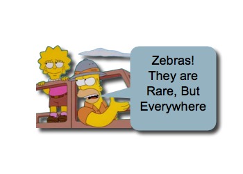 Hooven animals are complicated creatures (just like humans). They can be majestic, but rambunctious. They can be wild, yet tamed. In medicine, we often try to distinguish between them: Horses versus Zebras. While searching among the horses for the zebras, we may have in mind that the zebras are rare, which can be true on an individual basis; however, when the group you are searching through is large, the absolute number of zebras can be substantial (see Inborn Errors of Metabolism). The trick is to keep a vigilant eye open, trying to detect even the most subtle of stripes. One of those stripes that will catch your attention is ataxia. Let us take a moment to review one of the common “zebras” in children- Acute Cerebellar Ataxia:
Acute Cerebellar Ataxia: Basics
- Acute cerebellar ataxia is a common pediatric neurologic problem.
- Incidence of 1 in 100,000 – 500,000.
- Some causes of ataxia in children: [Thakkar, 2016]
- Post-infectious Cerebellar Ataxia – (~30 – 60%)
- Drug Intoxication (~8%)
- ex, Alcohol, Benzos, Heavy Metals, CO poisoning, Anticonvulsants
- Opsoclonus Myoclonus Ataxia (~8%)
- Rare, but a true medical emergency!
- May be misdiagnosed as benign post-infectious cause at first.
- Has severe ataxia, opsoclonus (chaotic ocular movements), and myoclonus.
- Is a Paraneoplastic disorder (often neuroblastoma)! [Tate, 2005]
- Acute Cerebellitis (~2%)
- Most severe end of the spectrum of cerebellar inflammation/infection. [Rossi, 2016]
- Previously, “Acute Cerebellitis” was used interchangeably with Post-infectious, But:
- Acute Cerebellitis has a distinctly worse disease course.
- Has abnormalities on brain MRI.
- Can lead to rapid posterior fossa edema and lead to morbidity and mortality.
- Cerebellar Stroke (~2%)
- Yes, stroke can occur in children.
- Look for risk factors, like:
- Acute Disseminated Encephalomyelitis (ADEM) (~2%)
- Immunologically mediated inflammatory disease
- Polyfocal neurological signs (multiple sites involved in CNS)
- Rapid onset of encephalopathy (altered mental status)
- Meningitis (<1%)
- Cerebral Venous Thrombosis (<1%)
- Miller Fisher Syndrome (<1%)
- Hereditary conditions (ex, Ataxia-telangiectasia)
Acute Cerebellar Ataxia: Post-infectious
- The most common cause of acute cerebellar ataxia in children is post-infectious cerebellar ataxia. [Thakkar, 2016; Rossi, 2016]
- Generally seen in kids younger than 6 years.
- Most common among 2 – 4 year olds.
- Presents in a relatively well appearing child who has: [Doan, 2016]
- Lack of coordination of movement NOT due to paresis,
- Alterations in tone,
- Sensory loss, and/or
- Involuntary movements
- Often, symptoms begin suddenly.
- NOT associated with fever, seizures, change in mental status, or other systemic signs. [Doan, 2016]
- Is a diagnosis of exclusion, because other ominous conditions can present similarly.
- Commonly associated infections:
- Varicella [Fursow, 2013]
- Chickenpox is frequently the cause of acute cerebellar ataxia in the immunosuppressed patient. [Fursow, 2013]
- Varicella vaccination, however, is protective. [van der Maas, 2009]
- Epstein-Barr virus
- Echovirus
- Enterovirus (Coxsackievirus)
- Varicella [Fursow, 2013]
- Work-up is generally negative!
- Cerebrospinal fluid analysis has low diagnostic yield. [Thakkar, 2016]
- Certainly CSF analysis is helpful if you are more concerned for meningitis or encephalitis.
- LP, if performed, should wait until after imaging to rule-out posterior fossa mass or edema. [Doan, 2016]
- Imaging is typically normal. [Thakkar, 2016; Doan, 2016]
- MRI is preferred given higher resolution and superior imaging of posterior fossa. [Rossi, 2016]
- CT should be obtained for patients with altered mental status, atypical disease course, asymmetric focal neurologic deficits, or when hemorrhage or mass is higher on the Ddx list.
- “Basic Labs” will be normal.
- Glucose is always worth checking!
- Electrolytes and urine catecholamines may be useful if concern for opsoclonus-myoclonus.
- Urine Tox screens should be considered, particularly in the toddlers who like to eat random items in the house. [Doan, 2016]
- Cerebrospinal fluid analysis has low diagnostic yield. [Thakkar, 2016]
- Patient recover without lasting sequelae. [Thakkar, 2016]
- Usually has resolution of symptoms in 2-8 weeks.
- Complete resolution by 2-3 months.
Moral of the Morsel:
- Zebras are common collectively! Look for the subtle stripes!
- Make kids walk! Yes, toddlers do “toddle,” but shouldn’t be ataxic!
- Look at the eyes! Nystagmus may be seen with benign conditions, but opsoclonus is scary!
References
Thakkar K1, Maricich SM2, Alper G3. Acute Ataxia in Childhood: 11-Year Experience at a Major Pediatric Neurology Referral Center. J Child Neurol. 2016 Aug;31(9):1156-60. PMID: 27071467. [PubMed] [Read by QxMD]
Rossi A1, Martinetti C2, Morana G2, Severino M2, Tortora D2. Neuroimaging of Infectious and Inflammatory Diseases of the Pediatric Cerebellum and Brainstem. Neuroimaging Clin N Am. 2016 Aug;26(3):471-87. PMID: 27423804. [PubMed] [Read by QxMD]
Doan TT1, Masom CP1, Mazzaccaro RJ2, Kane KE1. Acute Cerebellar Ataxia: An Unusual Pediatric Case. J Emerg Med. 2016 May;50(5):769-72. PMID: 26899517. [PubMed] [Read by QxMD]
Poretti A1, Benson JE, Huisman TA, Boltshauser E. Acute ataxia in children: approach to clinical presentation and role of additional investigations. Neuropediatrics. 2013 Jun;44(3):127-41. PMID: 23254568. [PubMed] [Read by QxMD]
van der Maas NA1, Bondt PE, de Melker H, Kemmeren JM. Acute cerebellar ataxia in the Netherlands: a study on the association with vaccinations and varicella zoster infection. Vaccine. 2009 Mar 18;27(13):1970-3. PMID: 19186201. [PubMed] [Read by QxMD]
Tate ED1, Allison TJ, Pranzatelli MR, Verhulst SJ. Neuroepidemiologic trends in 105 US cases of pediatric opsoclonus-myoclonus syndrome. J Pediatr Oncol Nurs. 2005 Jan-Feb;22(1):8-19. PMID: 15574722. [PubMed] [Read by QxMD]

[…] become easier for us to evaluate, as we encounter them commonly; however, the complaint of Vertigo, Ataxia, weakness, numbness, or visual changes can easily leave us scratching out heads. One item can come […]
Isd Acute Cerebellar Ataxia Hereditary ?
[…] of the leg – Appendicitis, PID, Epididymitis, Ovarian Torsion, Testicular Torsion, or Acute Cerebellar Ataxia. After an extensive review of your Ddx list, you may be inclined to call it “Growing […]
Pedantic point of order…The “Miller-Fisher Syndrome” you mention in this morsel is often erroneously hyphenated suggesting 2 contributing clinicians. In fact Dr Charles Miller Fisher, the neurologist after whom this variant of Guillain–Barré syndrome is named, had no hyphen between his names.
Pedantic, but proper.
Will be changed.
Gracias,
sean