![]()
Round Pneumonia
We have previously discussed Pediatric Pneumonia (and why I still live outside in the dog house). We all commonly assess the pediatric CXR and need to be skilled in its interpretation; but have you ever looked at a CXR of a child and asked yourself, “why is that mass so round? That has to be something more than an infection.” Well, for those of us who care of adults, that sick feeling you have in your gut that this round opacity is more than a pneumonia is often justified. Here, however, is another example of kids not playing by the same rules that adults do. Round Pneumonia is a well-known clinical entity in pediatrics, and fortunately, much more common than other “badness.”
Round Pneumonia – Interesting Anatomy
- Round pneumonia more commonly occurs in children.
- Usually seen in children under 8 years of age.
- Mean age 3-5 years.
- 75% under 8 years of age, 90% under 12 years of age.
- This is likely due to underdeveloped lung architecture.
- Underdeveloped Pores of Kohn
- Openings that are between adjacent alveoli.
- Interalveolar connections.
- Underdeveloped Channels of Lambert
- Channels that connect terminal bronchioles opening to alveolar sacs.
- Act as collateral circulation of gases.
- Both permit passage of fluid and bacteria, which allows the infection to spread more readily to the adjacent alveolar sacs.
- With underdeveloped connections, the infection cannot spread as easily and, therefore, has sharp margins and spreads outwardly in a spherical fashion.
- Underdeveloped Pores of Kohn
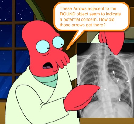
Round Pneumonia – Appearance
- Well-defined borders
- Most often solitary
- Located
- Posteriorly
- Lower Lobes
- Adjacent to fissures
Round Pneumonia – The Bugs
- Most commonly attributed to S. pneumoniae.
- Naturally, can also be caused by other bacteria and also viruses (SARS).
Round Pneumonia – Is it something else?
- Naturally, our job is to not to jump to conclusions and to keep our differential diagnosis list open… so what should you consider when looking at a Round Opacity?
- Lung Abscess (look real closely for any air-fluid levels)
- Fungal Infection
- TB
- Pulmonary anatomic malformations (ex, congenital cysts, sequestration)
- Neoplasms (ex, lymphoma, neuroblastoma, solid tumors)
- Diaphragmatic Hernia
Round Pneumonia – When to Consider CT
In contrast to older kids and adults, a round opacity in a child is much more likely to be a pneumonia than a malignancy. With this in mind, and with a desire to utilize CT scans judiciously, it is generally best to treat Round Pneumonia with antibiotics and save the CT for those kids who:
- have clinical features that are NOT consistent with pneumonia,
- have a round opacity that does NOT resolve after antibiotics,
- have radiographic evidence signs of nonpulmonary origin on CXR (ex, Calcifications, bony destruction).
Liu YL, Wu PS, Tsai LP, Tsai WH. Pediatric Round Pneumonia. Pediatrics and Neonatology. 2013. http://dx.doi.org/ 10.1016/j.pedneo.2013.01.014
Restrepo R, Palani R, Matapathi UM, Wu YY. Imaging of Round Pneumonia and Mimics in Children. Pediatr Radiol. 2010; 40: 1931 – 1940.
Kim YW, Donnelly LF. Round Pneumonia: Imaging Findings in a Large Series of Children. Pediatr Radiol. 2007; 37: 1235 – 1240.

[…] special attention. We have discussed pneumonia several times previously (ex, Pneumonia Detective, Round Pneumonia, Penicillin for Pneumonia, and CAP), but recently our friends at the Section on Emergency […]
[…] Fox takes us through recognising and managing round pneumonias in Pediatric EM Morsels. […]
[…] Round Pneumonia […]