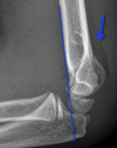
Supracondylar Fracture
It is August… a few weeks left before school starts… and football season is almost upon us! I love football season, but it is such a double-edged sword – inevitably all of the Peds EDs will be inundated with skeletally immature gladiators having injured themselves. So prepare yourself for the onslaught of supracondylar fractures!
Supracondylar Fractures
- The supracondylar area is composed of thin, weak bone.
- Supracondylar fx’s account for >50% of all pediatric fx’s.
- Peak incidence is 5-7 years.
- Two main types based on mechanism
- Extension-type (FOOSH)
- Most common (~95%)
- Leads to posterior displacement of the fracture
- Flexion-type
- From a direct blow to the posterior aspect of the elbow while it is flexed
- Leads to anterior displacement of the fracture
- Extension-type (FOOSH)
- Three main classifications
- Type I – non-displaced, limited radiographic evidence of fx
- Type II – angulated and displaced, but still partially attached
- Type III – complete displacement without any connection
Evaluation
- Evaluation (and Documentation!!) of the Vascular and Neurologic status of the affected arm is imperative!
- Supracondylar Fx have a great potential for nerve and/or vascular compromise.
- Vascular
- Brachial artery may be injured with posteriorly displaced fxs.
- Documenting radial and ulnar pulses is good… BUT
- Absence of brisk cap refill is the best indicator of long-term vascular injury.
- The patient with diminished pulses who has brisk cap refill will likely do well after reduction that is done in an urgent fashion.
- The patient with diminished pulses and who has poor distal perfusion needs emergent intervention!
- Using pulse oximetry can also help document peripheral perfusion in the affected arm, if it there is a good waveform and oxygen saturation.
- Motor Nerve
- Often getting the child with a deformed arm to cooperate for examination is difficult… give them Pain Meds!!
- Quick and Dirty nerve exam of the arm
- Thumb’s up = Radial Nerve
- Spread fingers out (“like Michael Jordan palming a basketball”) = Ulnar Nerve
- Abduction of the Thumb (“OK sign”) = Median Nerve
- Sensory Nerve
- Dorsal aspect of 1st web space = Radial Nerve
- Palmer aspect of pinky = Ulnar Nerve
- Palmar aspect of pointer finger = Median Nerve
Consider Complications
- Always consider Compartment Syndrome
- Supracondylar Fx are at high risk for it
- Assess, reassess, and reassess again
- Look for Pain, Pallor, Pulselessness, Paralysis, and Paresthias
- Pain with passive range of motion of distal fingers is very concerning!!
Subtle radiographic findings
- The Type II and Type III fractures are often no-brainers… bone looks broke!
- Type I can be very subtle… so look for:
- Displaced Anterior Fat Pad (Sail Sign)
- Any Posterior Fat Pad
- Anterior Humeral Line not intersecting the middle third of the capitellum.
- Radiocapitellar line not interesting the middle third of the capitellum.
Shrader MW. Pediatric supracondylar fractures and pediatric physeal elbow fractures. Orthop Clin N Am. 2008; 39:163-171.
Sarraff LM, Haines CJ. Common orthopedic injuries in the pediatric ED. Pediatric Emergency Medicine Reports. 2010; 15 (7): 77-92.


[…] aware that even fractures deserve more attention than describing what bone is broken (ex, wrist, supracondylar, pelvic, toddler’s). Unfortunately, we have to contemplate the potential for a patient […]
[…] manage are the more common orthopedic varieties (ex, Patellar Dislocation, Shoulder Dislocation, Supracondylar Fracture). Unfortunately, while we all want/need our children to be active, this activity may lead to some […]
[…] orthopaedic topics previous in the PedEM Morsels (ex, osteomyelitis, patellar dislocation, SCFE, supracondylar fractures, nursemaid’s elbow), but let’s take a moment to look at yet another: Shoulder […]