![]()
Ear Foreign Body
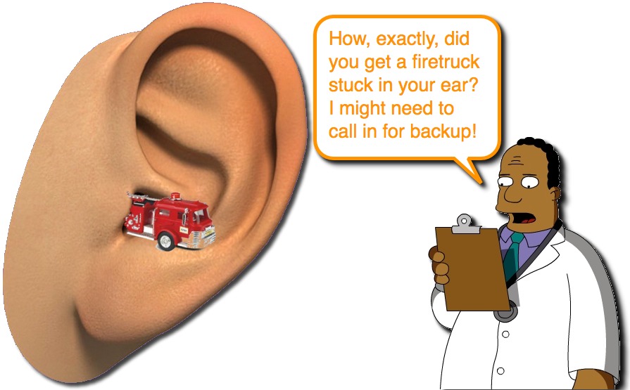
Without question, kids will put the oddest objects into their orifices… and then say that they don’t know how they got there. While this can lead to same comic relief to your otherwise stressful shift, managing a foreign body can also generate its own level of stress. We have great hopes for the offending foreign body being easy to remove as we walk into the room, but then can be derailed by a tenacious monster that refuses to leave and a wailing and flailing child. Those don’t make for any comic relief.
We have discussed many topics with respect to the pediatric foreign body (Aspirated FB, Nasal FB, Button Batteries in the nose, Esophageal Button Batteries), but let us look at the other common entity – the Ear Foreign Body.
Anatomy Matters
- The anatomy of the ear seems simple… an external portion, a tube, and the ear drum. Naturally, it is more complicated than that.
- The External Auditory Meatus can be divided into two portions:
- The lateral third – cartilaginous portion.
- The medial two thirds
- Bony
- More narrow than the lateral third.
- Lined with a very vascular and highly sensitive thin layer of skin.
- Prone to bleeding with even slight trauma.
Position Matters
- Naturally, the position of the patient can affect your ability to successfully remove the foreign body.
- Make sure that the patient is in a comfortable position for him/her.
- Sitting on a parent’s lap, sideways with the ear in question easily visible will often be the first position of choice.
- This will allow the patient’s legs to be between the parent’s and offer security.
- Also allows for the torso to be supported and arms kept safely out of the way.
- Teaching the family member how to do this can save everyone some time and sweat.
- Sitting on a parent’s lap, sideways with the ear in question easily visible will often be the first position of choice.
- Equally important is to make sure that the patient is in a position that allows you to be comfortable.
- You may be very good a yoga, but there is no need to demonstrate your skills while attempting to remove a foreign body.
- Anticipate that this will not be easy and may take a few minutes… so position your tools within easy reach.
- Make sure that the patient is in a comfortable position for him/her.
- The position of the Foreign Body is also very important.
- Foreign bodies that are in the medial two thirds of the canal are much more problematic and more difficult to remove.
- In this area, the patient will be more uncomfortable and less likely to hold still.
- You are also more apt to cause trauma (to the canal or TB) with foreign bodies that are in this region.
- You need to have optimal circumstances and be very careful with foreign bodies in the medial two thirds of the canal.
Pain Management Matters
- Even the most cooperative child will loose the ability to calmly sit still as you scrap and claw at a foreign body, especially one in the medial two thirds of the canal.
- Be kind. Use some pain medications when appropriate.
- A common trick is to use topic lidocaine.
- It is important to first make sure that there is no perforation of the TM before pouring any fluids into the canal.
- This is very helpful with insects that are entrapped, as it will drown the insect and make everyone’s job easier.
- Ketamine is great too!
- Ok, so this isn’t something to pull out right away, but occasionally, the child with an ear foreign body will need procedural sedation.
- Short acting agents would be ideal (propofol). ENT would likely go to the OR for anesthetic gas.
The Object Matters
- A endless variety of foreign bodies have been removed from ears (from cotton to cheese; from bead to popcorn kernel; from eraser to putty).
- There are characteristics that can make the Foreign Body more difficult to deal with:
- Vegetable Matter (food, beans, etc)
- Do not use irrigation as this may cause the foreign body to swell and become more entrapped.
- Button batteries
- Cause liquefaction necrosis and need to be removed promptly.
- Sharp objects
- If not removed very carefully, may cause more injury and damage during the removal.
- Smooth, round objects
- Difficult to grasp.
- Especially challenging when they are in the medial 2/3 of the canal.
- Vegetable Matter (food, beans, etc)
The Tool Matters
- Having a wide array of tools can help you adapt to the various challenges that each foreign body offers.
- These can come in handy:
- Magnet
- Forceps (alligator and Hartman)
- Frazier suction
- Cerumen loop
- Right-angle ball hook
- Schuknecht foreign body remover
- Aural irrigation devise
- Can be made using a 60 ml syringe and an 18 gauge angiocath.
- Ensure that the water is body temperature (so you don’t make the child vertiginous).
- Otomicroscope would be quite handy.
Knowing Your Limits Matters
- Occasionally, you won’t be successful.
- When should you refer to the ENT doctors?
- Unable to remove after multiple attempts.
- Multiple attempts increases risk of complications, which can lead to other long term issues.
- Tightly wedged objects
- Objects resting against the TM
- Sharp objects
- Button batteries
- These, however, cannot wait until the next day in the ENT office.
- If you can’t get it out, then the ENT needs to come in to get it out.
- Unable to remove after multiple attempts.
Reexamination Matters
- After you have successfully removed the Foreign Body, your job is not done.
- Ensure that all parts have been removed.
- Especially important for insects.
- Residual barbed insect legs can lead to inflammation and damage.
- Ensure that no other orifice has a Foreign Body.
- Kids are tricky.
- Check their other ear and the nostrils!!
Post-Care Matters
- Important to educate the patient and family about the hazard of the foreign body (this time it was the ear, next time it might be the airway).
- If there was some trauma to the canal (laceration, inflammation), then prescribe antibiotic otic drops and give water precautions. Follow-up examination will be important.
References
Stoner MJ1, Dulaurier M. Pediatric ENT emergencies. Emerg Med Clin North Am. 2013 Aug;31(3):795-808. PMID: 23915604. [PubMed] [Read by QxMD]
Cederberg CA1, Kerschner JE. Otomicroscope in the emergency department management of pediatric ear foreign bodies. Int J Pediatr Otorhinolaryngol. 2009 Apr;73(4):589-91. PMID: 19168230. [PubMed] [Read by QxMD]
Brown L1, Denmark TK, Wittlake WA, Vargas EJ, Watson T, Crabb JW. Procedural sedation use in the ED: management of pediatric ear and nose foreign bodies. Am J Emerg Med. 2004 Jul;22(4):310-4. PMID: 15258875. [PubMed] [Read by QxMD]
DiMuzio J Jr1, Deschler DG. Emergency department management of foreign bodies of the external ear canal in children. Otol Neurotol. 2002 Jul;23(4):473-5. PMID: 12170148. [PubMed] [Read by QxMD]
Schulze SL1, Kerschner J, Beste D. Pediatric external auditory canal foreign bodies: a review of 698 cases. Otolaryngol Head Neck Surg. 2002 Jul;127(1):73-8. PMID: 12161734. [PubMed] [Read by QxMD]
Ansley JF1, Cunningham MJ. Treatment of aural foreign bodies in children. Pediatrics. 1998 Apr;101(4 Pt 1):638-41. PMID: 9521948. [PubMed] [Read by QxMD]
Author

[…] They are the focus of many ED visits! Things get stuck in them (ex, Ear Canal Foreign Body, Cerumen). They can become inflamed from swimming (ex, Swimmer’s Ear). Infection, though, is […]
[…] we encounter in the Emergency Department. Many make you scratch your head (how did they get that FB in the Ear?) and others will challenge you (how do I get this fish hook out of the kid’s face?), but one […]
[…] we attribute to exploring and learning about the world (ex, putting objects in nostrils, ears, or mouth). Other times the “odd conditions” are not quite explained, but are well […]
[…] previous Ped EM Morsels have discussed several foreign body topics (ex, Aspirated FB, Ear FB, Delayed Dx of Aspirated FB, Button Battery FB, Nasal FB). There is another type of foreign […]