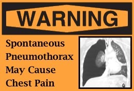
Spontaneous Pneumothorax
Spontaneous Pneumothorax
We all see a myriad of patients who complain of chest pain and/or shortness of breath. We can rapidly develop the differential diagnosis and deftly sort through it without much effort; however, we often still end with the question of whether to order a CXR. I commonly will justify getting a CXR by stating that a spontaneous pneumothorax could be present. Yes, I know it is rare, but it is a needle in the haystack of disease that warrants consideration.
Basics
- Spontaneous pneumothorax (SPtx) is uncommon… it occurs more commonly in adults than children.
- Most recent data: incidence = 2.6 cases per 100,000
- Primary vs Secondary SPtx
- Etiology of SPtx is, bascially, due to connective tissue alterations that predispose to leakage of air from the airways into the pleural space.
- Primary SPtx– occurs in the absence of known underlying lung condition
- There seems to be a subset of Familial Primary SPtx – so Family Medical History is important.
- Seconday SPtx– occurs as a consequence of underlying lung pathology
- Asthma, Cystic Fibrosis, Emphysema
- Marfan Syndrome, Ehlers-Danlos, Lupus, other connective tissue disorders
- Infections
- Malignancies (ex, lymphoma)
- Foreign Bodies
- Congenital malformation
- Catamenial (associated with menses)
- Secondary SPtx can occur in patient with history of asthma WITHOUT having an asthma exacerbation.
Who is at risk?
- Kids 10-17 years of age are at greatest risk (~94% of SPtx occurred in kids 10 yrs and older).
- Male predominance (~80% of cases are males).
- History of Asthma or tobacco use increases risk.
Presentation
- Most will have Acute Onset of Chest Pain and/or Shortness of Breath
- While actions that increase intrathoracic pressure (lifting, straining, Valsalva, etc) can precipitate SPtx, it often occurs while at rest.
- Patients may actually present in a delayed fashion and note slowly resolving symptoms over the past 1-3 days, but still have a SPtx.
- Examination may be abnormal (decreased breath sounds, symmetric chest rise, etc); however, small Ptx’s or Ptx’s in smaller children may produce no identifiable physical exam abnormalities.
- Minority of patients (6.6% of all SPtx cases) will present with tension physiology (ideally, we’d identify that on examination).
Diagnosis
- Chest X-Ray
- Upright CXR is most common method to diagnose
- Can do Decubitus Views to increase sensitivity
- CT
- Superior to CXR; however, does that increased sensitivity outweigh the radiation risk (see Radiation Morsel)?
- CT may be warranted in the work-up of the patient with known pneumothorax to investigate for Secondary Etiology (ex, lung mass) and risk stratify for recurrence (Blebs detected on the contra-lateral side may portend future occurrences).
- Best to discuss the utility of CT with the surgeon on a case by case basis.
- Ultrasound
- There are plenty of studies that demonstrate that U/S is more sensitive than supine CXR in the setting of adult trauma.
- U/S – ~90% sensitive
- Supine CXR – ~50% sensitive
- Erect CXR has increased sensitivity (~90%), naturally.
- U/S is naturally operator dependent… and in this case the operator is you… so are you dependable?
- This is a quick basic review of thoracic ultrasound.
- Look here for a quick demonstration by Dr. Tony Weekes.
Change My Practice?
My humble opinion: For the child that presents with chest pain and/or dypnea you should consider pneumothorax, but that doesn’t necessarily mean you need to obtain a chest xray on all of them. If the story is concerning (ex, acute onset, persistent, associated with straining/valsalva) then you need to image. Your threshold to image should be lower for those who have history of asthma, use tobacco, have a family history of SPtx, or are older than 10 years of age.
How should you image? Well, I would advocate for starting with bedside ultrasound (if you are savvy). It has essentially the same sensitivity as erect CXR and has several advantages.
- It doesn’t require the child to wait for radiology.
- The kids, particularly the older kids, think it is cool … which earns you patient satisfaction points.
- There is no radiation, and you can “sell” that to the parents and earn even more satisfaction points.
- When you are done, you have an answer and that delivers even more satisfaction points!
If U/S is not an option for you… Erect CXR is the answer. Save CT for pts who have increased risk for other pathology (PE, malignancy, etc).
Management
- Many patients can be managed without intervention (small pneumothorax, minimal symptoms).
- If you are going to do an intervention, consider the most minimally invasive approach first (see procedure videos).
- Needle aspiration
- Pigtail Catheter
- Chest Tube
Dotson K, Timm N, Gittelman M. Is Spontaneous Pneumothorax Really a Pediatric Problem? A National Perspective. Pediatric Emer Care 2012; 28(4): 340-344. Dotson K, Johnson LH. Pediatric Spontaneous Pneumothorax. Pediatric Emer Care 2012; 28(7): 715-723.


Is it possible that a pneumothorax doesn’t show up on CXR? My 16 yr old son has the symptoms of pneumothorax, but no signs on the x-Ray. The Dr said she might have heard less intake on his left lung but nothing major. I had 2 pneumothoraxes in my twenties and my sister had one I her late twenties.
Jackie,
Yes, it is possible to have a pneumothorax and not see it on an Xray… although, these tend to be smaller and less consequential. Sometimes additional X-rays can be taken, or an Ultrasound used.
-sean
[…] conditions and complications. Previously, we have discussed abnormal collections of air (ex, pneumothorax, pneumomediastinum). Now, let us look at another potentially alarming situation- Pneumatosis […]