![]()
Proximal Tibiofibular Joint Dislocation in Children
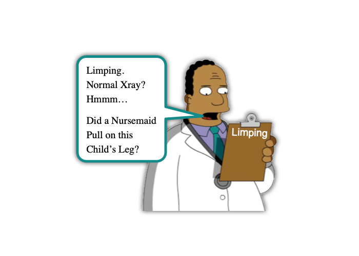
In the past, we have discussed several causes of Knee Pain in children. Some of those causes are benign (ex, Osgood Schlatter’s Disease) while others are scary (ex, osteosarcoma). Evaluating the limping child, though, requires us to ponder not only the common (ex, Toddler’s Fracture), but also to be vigilant for the severe (ex, Septic Arthritis). Let’s take a moment to digest Morsel that pertains to another interesting cause of knee pain and limping in a child. This one can be thought of as being akin to the radial head subluxation (ie, Nursemaid’s Elbow) of the knee – Proximal Tibiofibular Joint Dislocation:
Proximal Tibiofibular Joint Dislocation in Children: Basics
- Proximal Tibiofibular Joint (PTF) dislocations are relatively rare. [Cunningham, 2018]
- Account for less than 1% of all knee injuries.
- The PTF joint is inherently stable with good ligamentous support, especially when the knee is in extension. [Horan, 2006]
- Anatomy of the PTF Joint [Cunningham, 2019]
- The PTF joint is a synovial joint between the lateral condyle of the tibia and the head of the fibula.
- It is reinforced with an articular capsule.
- It is strengthened by the anterior and posterior tibiofibular ligaments.
- Mechanism of dislocation: [Horan, 2006]
- Typically, the injury occurs when:
- The Knee is in Flexion and
- The Foot is rotated and plantar flexed.
- Seen in sports that involved aggressive twisting of the knee (ex, snowboarding, soccer, horse-riding) as well as direct trauma.
- Typically, the injury occurs when:
- 4 Injury Patterns Described: [Cunningham, 2019; Horan, 2006]
- Subluxation (Type 1)
- Usually atraumatic
- Usually preadolescent patients
- Cases have occurred from simple acts like removing infant from car seat.
- The immature shape of the fibular epiphysis in young children may promote subluxation with minimal force. (Fortunately, this allows for reduction to also occur with minimal force … These are the ones that behave similar to radial head dislocation) [Sacchetti, 2022]
- Anterolateral Dislocation (Type 2)
- Most common (85%)
- Fall with knee flexed, ankle inverted, and foot plantar flexed
- Posteromedial Dislocation (Type 3)
- ~10% of cases
- Direct trauma (ex, impact from car’s bumper)
- Associated with Peroneal Nerve Injury
- Superior Dislocation (Type 4)
- 2% of cases
- Associated with High Energy ankle injuries and common peroneal injury
- Subluxation (Type 1)
Proximal Tibiofibular Joint Dislocation in Children: Management
- Diagnosis is primarily clinical [Horan, 2006]
- Having high index of suspicion is important.
- Key feature is lateral knee pain worsened by palpation over the fibular head.
- The rest of the exam may be completely normal other than discomfort with bearing weight.
- May have:
- Pain with knee extension
- Visual deformity
- Crepitance
- Locking or popping
- In Children, should consider it when: [Sacchetti, 2022]
- Patient refuses to bear weight,
- Has Painless Range of Motion of the Knee, and
- Tenderness over the Fibular Head.
- Plain films may be normal if subluxation is subtle… or may clearly show dislocation. [Horan, 2006]
- Normal AP knee film shows the fibular head overlapping the lateral margin of the lateral tibial condyle.
- Normal lateral knee film shows majority of the fibular head located posterior to the posterior margin of the tibia.
- Some cases do require CT or MRI to confirm. [Cunningham, 2019]
- Treatment [Horan, 2006]
- 1st option for Subluxation and Anterolateral Dislocation is Manual Reduction (similar to reducing that Nursemaid’s Elbow).
- MANAGE PAIN!
- Local anesthetic may be used.
- Procedural sedation may also be needed.
- Hold knee in flexed position at 90 degrees.
- Invert and Plantar Flex the ankle.
- Apply Downward pressure over the fibular head while extending the knee.
- A “pop” can be heard/felt.
- This is successful in most cases. [Cunningham, 2019]
- Then immobilize the joint and arrange follow-up.
- MANAGE PAIN!
- Posteromedial and Superior dislocations and failed manual reductions require open reduction and internal fixation.
- 1st option for Subluxation and Anterolateral Dislocation is Manual Reduction (similar to reducing that Nursemaid’s Elbow).
Moral of the Morsel
- Limping may be from more than septic arthritis! The Ddx list for Limping is broad… and now you have one more item to consider!
- Anatomy and Age Matters! The young patient’s fibular head requires less force to sublux… similar to the radial head!
- Another Mr. Miyagi Moment!? With appropriate pain control/sedation, you may be able to manually reduce the tibiofibular joint dislocation and heal with your bare hands!
References:
Sacchetti A, Verbaro J. Proximal tibiofibular joint dislocation in young children: Is this the nursemaid’s elbow of the lower extremity? Am J Emerg Med. 2022 Apr;54:328.e3-328.e4. doi: 10.1016/j.ajem.2021.10.054. Epub 2021 Nov 2. PMID: 34774384.
Cunningham NJ, Farebrother N, Miles J. Review article: Isolated proximal tibiofibular joint dislocation. Emerg Med Australas. 2019 Apr;31(2):156-162. doi: 10.1111/1742-6723.12989. Epub 2018 Apr 19. PMID: 29671944.
Horan J, Quin G. Proximal tibiofibular dislocation. Emerg Med J. 2006 May;23(5):e33. doi: 10.1136/emj.2005.032144. PMID: 16627827; PMCID: PMC2564106.
Author

I appreciate the article’s discussion of the different treatment options available. There are a variety of treatment options available, and the best option for each child will vary depending on the severity of the dislocation.