![]()
Pediatric Facial Fractures & Age
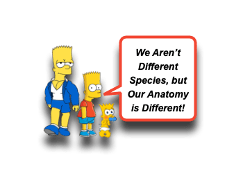
Often clinicians shy away from caring for children, because of the notion that “they aren’t little adults.” We have previously discussed my disdain for this mantra. They are not aliens! They do, however, have different anatomy and physiology that must be taken into consideration. What makes children even more exciting (AKA challenging) to care for is that these differences evolve and change as they grow older. We see this most obviously with respect to Trauma. Take facial trauma for example. Nasal fractures in kids need to be approached differently than in adults. Actually, all of the changing anatomy of a child affects facial fractures considerably. Let us take a minute to digest a morsel of knowledge on Pediatric Facial Fractures and Age:
Facial Fractures: Basics
- Facial Fractures occur less commonly in children than adults.
- ~5-15% of all facial fractures are seen in children. [Alcala-Galiano, 2008]
- Lowest rates in infants and increases with age.
- Two peaks in frequency of facial fractures: [Alcala-Galiano, 2008]
- 6-7 years of age (early school age)
- 12-14 years of age (increased sports participation… and being adolescents)
- Most common associated causes: [Alcala-Galiano, 2008]
- Motor Vehicle Accidents – ~36% (because cars injure everyone all of the time!) [Wong, 2016; Costa Ferreira, 2016]
- Sports Participation – ~26% (Kids colliding at high speeds, or throwing projectiles at each other)
- Falls – ~23% (Gravity works always) [Oleck, 2019]
- Interpersonal Violence – ~9% (’cause people are terrible to each other) [Hoppe, 2015; Hope, 2014]
- CHILD ABUSE should always be considered!
- As well as Bullying and Harassment
- Think beyond the injury… you may positively affect the kid’s future today!
- Associated Injuries: [Alcala-Galiano, 2008]
- Facial fractures are often associated with severe traumatic mechanisms. [Costa Ferreira, 2016]
- As much as 88% will have associated injuries. [Alcala-Galiano, 2008]
- Mid-face fractures can be associated with intra-cranial injury and cervical spine injury. [Hoppe, 2014]
- Increased risk of associated injuries depends on type of facial fracture:
- More complex fractures
- Midfacial and Mandibular fractures
- Fracture of “Protected Bone”
Facial Fractures: Changing Anatomy
- Fracture patterns in children are similar to adults, BUT likelihood of specific injury patterns change due to anatomic factors.
- There are 4 evolving anatomic factors that influence facial fracture frequency. [Alcala-Galiano, 2008]
- Skull-to-Face ratio
- Very young have prominent frontal skull protrusion with smaller facial features.
- Frontal skull often “protects” the face from impact.
- With increasing age, the face grows downward and forward, leading to the mid-face and mandible being the most prominent structures.
- From birth to maturity, the cranium increases in size 4-fold, but the face increased 12-fold! [Alcala-Galiano, 2008]
- Sinus Development
- Pneumatization of the sinuses is an important factor. [Alcala-Galiano, 2008]
- It progresses sequentially from the Ethmoids to the Maxillary sinuses, Sphenoid Sinuses, and ends with Frontal Sinuses.
- Prior to pneumatization, the bone is more resistant to fracture.
- After sinus development, the bone may fracture more easily, but that dissipates force (ie, cushions).
- Maxillary and Frontal Sinus development are correlated with frequency of mid-face fractures.
- Phase of Dentition
- Dentition occurs in 3 stages.
- Decidual phase (~ 2 years)
- Mixed phase (6-12 years)
- Permanent phase (>~12 years)
- The un-erupted teeth (during decimal or mixed phase) add strength to the maxillary and mandible. [Alcala-Galiano, 2008]
- Dentition occurs in 3 stages.
- Structure of Bone and Soft Tissues
- Bones in children are more elastic and resistant to fracture than adults.
- Higher likelihood of greenstick fractures in children. [Alcala-Galiano, 2008]
- The elasticity of the surrounding soft tissues can lead to minimal external signs of injury. [Oleck, 2019]
Facial Fractures: Types
- Nasal Fractures are most common – ~59% [Alcala-Galiano, 2008]
- Mandibular Fractures – ~21% (See Morsel) [Alcala-Galiano, 2008]
- Orbital – 9% [Alcala-Galiano, 2008]
- Rare in young children.
- Once frontal sinus develops (~ 7 yrs), force is transmitted to the medial and lateral walls and floor of the orbit.
- Orbital Fractures are often associated with other fractures! [Oleck, 2019]
- Common fracture to look for is the Orbital Blowout fracture!
- Floor is the least resistant bone.
- May see enophthalmos.
- Patient may have diplopia.
- Look for Ocular injuries!
- Frontal Skull – ~5% [Alcala-Galiano, 2008]
- More common in younger children due to prominence of forehead.
- In kids < ~7 yrs (frontal sinus has not developed), frontal fractures tend to affect the orbital roof!
- Considered to be Skull Fractures at this age.
- Increased risk for neurocranial injuries. [Alcala-Galiano, 2008]
- Midface – ~3% [Alcala-Galiano, 2008]
- Rare in young children.
- Typically result from high-energy impacts.
- Prevalence increases as maxillary sinuses develop and permanent teeth erupt.
- Zymgomatic fractures are the most frequent fractures of the mid-face, but often greenstick fractures in children.
- Can involve the orbital floor and walls.
- Complex Naso-orbital (ie, Le Fort) [Alcala-Galiano, 2008]
- Are least common – ~1%
- Greatest potential for future deformity.
Moral of the Morsel
- In the beginning, the Face is Protected. The large forehead will often take the brunt of the force.
- With age, comes a Bigger Face! As the sinuses develop and the teeth erupt, and the face grows, it becomes more at risk.
- Don’t just look for the fracture! There may be more associated injuries!
- Don’t just stop with the injury. Ask why? Inter-personal violence is a problem that warrants our action.
References
Oleck NC, Dobitsch AA, Liu FC, Halsey JN, Le TT, Hoppe IC1, Lee ES, Granick MS. Traumatic Falls in the Pediatric Population: Facial Fracture Patterns Observed in a Leading Cause of Childhood Injury. Ann Plast Surg. 2019 Apr;82(4S Suppl 3):S195-S198. PMID: 30730318. [PubMed] [Read by QxMD]
Reich W1, Aust O2, Eckert A3. Prospective analysis of mid-facial fractures in a single-center pediatric-adolescent cohort. Int J Pediatr Otorhinolaryngol. 2019 Apr;119:151-160. PMID: 30708183. [PubMed] [Read by QxMD]
Lee SH1, Yun SJ2, Ryu S1, Choi SW1, Kim HJ1, Kang TK1, Oh SC1, Cho SJ1. Brain Computed Tomography Compared with Facial 3-Dimensional Computed Tomography for Diagnosis of Facial Fractures. J Pediatr. 2017 May;184:32-37. PMID: 28190518. [PubMed] [Read by QxMD]
Ferreira PC1, Barbosa J, Braga JM, Rodrigues A, Silva ÁC, Amarante JM. Pediatric Facial Fractures: A Review of 2071 Fractures. Ann Plast Surg. 2016 Jan;77(1):54-60. PMID: 25275475. [PubMed] [Read by QxMD]
Wong FK1, Adams S, Coates TJ, Hudson DA. Pediatric Facial Fractures. J Craniofac Surg. 2016 Jan;27(1):128-30. PMID: 26674891. [PubMed] [Read by QxMD]
Hoppe IC1, Kordahi AM, Lee ES, Granick MS. Pediatric Facial Fractures: Interpersonal Violence as a Mechanism of Injury. J Craniofac Surg. 2015 Jul;26(5):1446-9. PMID: 26106996. [PubMed] [Read by QxMD]
Hoppe IC1, Kordahi AM, Paik AM, Lee ES, Granick MS. Examination of life-threatening injuries in 431 pediatric facial fractures at a level 1 trauma center. J Craniofac Surg. 2014 Sep;25(5):1825-8. PMID: 25203578. [PubMed] [Read by QxMD]
Iso-Kungas P1, Törnwall J, Suominen AL, Lindqvist C, Thorén H. Dental injuries in pediatric patients with facial fractures are frequent and severe. J Oral Maxillofac Surg. 2012 Feb;70(2):396-400. PMID: 22260909. [PubMed] [Read by QxMD]
Imahara SD1, Hopper RA, Wang J, Rivara FP, Klein MB. Patterns and outcomes of pediatric facial fractures in the United States: a survey of the National Trauma Data Bank. J Am Coll Surg. 2008 Nov;207(5):710-6. PMID: 18954784. [PubMed] [Read by QxMD]
Alcalá-Galiano A1, Arribas-García IJ, Martín-Pérez MA, Romance A, Montalvo-Moreno JJ, Juncos JM. Pediatric facial fractures: children are not just small adults. Radiographics. 2008 Mar-Apr;28(2):441-61; quiz 618. PMID: 18349450. [PubMed] [Read by QxMD]
Author
