![]()
Facial Hemangioma
The skin is the largest organ, yet we often undervalue its importance. Ok, maybe it is just me being less than comfortable with all of the oddities that can present with skin findings (ex, pyoderma gangrenosum). This is why I approach rashes the way I do (see, Rash). Certainly, there are conditions that warrant greater concern (ex, Kawasaki Disease Shock Syndrome, Erythema Multiforme, RMSF, Eczema Herpeticum), but fortunately, many skin eruptions are just annoying (ex, Mulluscum). There is one skin finding that can exist anywhere on this spectrum though: it may be a simple and self-resolving condition, or just the tip of the iceberg. Let’s take a minute to review the Facial Infantile Hemangioma:
Facial Hemangioma: Hemangioma vs Vascular Malformation
Hemangiomas and Vascular Malformations (see AVM) are not the same entities. [Darrow, 2015; Williams, 2000]
- Hemangiomas:
- True neoplasm, histologically, with increased cellular turnover.
- Do not undergo malignant transformation, but…
- Can lead to complications
- Start small, but… have a Proliferative Phase
- May be present at birth, but often only seen as a small, red macule or localized telangiectasia.
- Usually present in first few weeks of life (in first 8 weeks of life).
- Proliferative, growth continues through 8-12 months.
- Functional complications can develop during this period.
- Involution phase follows after proliferation phase
- Tumor rate of growth is proportionally less than growth rate of the child.
- Tumor becomes less firm.
- Vascularity replaced by fibro-fatty tissue.
- Color changes from bright, shiny red to grayish and mottled.
- Involution continues over the next 1 – 12 years of childhood – most by 4yrs.
- True neoplasm, histologically, with increased cellular turnover.
- Vascular Malformations:
- NOT true neoplasm, but are errors of vascular morphogenesis
- Can be Capillary, Venous, Arterial, Lymphatic
- Can also be a Combination of 2 or more types
- Present at birth
- Increase in size throughout life
- Growth in size depends on intraluminal hydrostatic pressures.
- Based on pressures, some increase more rapidly than others
- Do NOT involute.
- NOT true neoplasm, but are errors of vascular morphogenesis
Differentiating between the two entities is important as prognosis, complications, and therapies are different. [Williams, 2000]
Hemangioma Therapies
- Challenge of managing hemangiomas in children is in trying to predict whether they will cause complications before they spontaneous involute and whether that risk favors treatment. [Williams, 2000]
- Management is based on size/depth, location, and potential for complication. [Darrow, 2015; Min-Jung, 2013; Williams, 2000]
- There are several management strategies that are used: [Darrow, 2015; Williams, 2000]
- Beta-Adrenergic Blockers (ex, Propranolol)
- Often used as first line medical therapy. [Darrow, 2015]
- Superficial hemangiomas may respond to topical timolol.
- Robust evidence is still lacking.
- Steroids
- Hemangiomas respond to systemic and intralesional steroids.
- Systemic steroids given for “croup” can temporarily improve subglottic hemangiomas also. (see below)
- Pulse-Dye Laser
- Wavelength of 577 nm affects hemoglobin and leads to lysis of the vascular lesion while sparing surrounding tissues.
- Pulse width of 450 microseconds helps avoid thermal injury as well.
- Only helpful for superficial or dermal hemangiomas (< 1mm).
- Surgical Excision
- Used when less invasive therapies have not been effective.
- Used when focal involvement threatens adjacent anatomic structures.
- Beta-Adrenergic Blockers (ex, Propranolol)
Hemangioma: Complications
- Facial hemangiomas more frequently cause complications than non-facial hemangiomas.
- Predictors of complications: [Haggstrom, 2006]
- Large size
- Facial location
- Segmental morphology
- Common Types of Complications: [Haggstrom, 2006]
- Local / adjacent structure compression
- Visual loss
- Respiratory distress / airway obstruction
- Cartilaginous destruction (ex, nose)
- Ulceration
- Bleeding
- Congestive heart failure
- Scarring and cosmetic disfigurement
- Local / adjacent structure compression
- BONUS Morsel: The presence of more than 5 focal infantile hemangiomas suggests higher risk for hepatic involvement. [Darrow, 2015]
Hemangioma: “Special” Locations
- Periorbital [Darrow, 2015]
- Hemangiomas of the periocular area have the potential to compress the globe or local structures.
- May cause refractive errors, strabismus, amblyopia, and visual loss.
- High risk hemangiomas are those >1cm in diameter, located near the nose, associated with ptosis or eyelid margin change, or globe displacement.
- Subglottic [Darrow, 2015; O-Lee, 2008]
- May be associated with concurrent hemangioma in the “Beard Distribtion” – especially multiple hemangiomas in this distribution.
- Most common location of airway infantile hemangioma.
- Causes biphasic (inspiratory and expiratory) stridor!
- Can be confused with Croup… but … won’t have fever or rhinorrhea.
- If treated with oral steroids (for croup), may improve, but will recur. (Remember, recurrent Croup… may not be croup).
- Severe obstruction may require excision or tracheostomy.
Moral of the Morsel
- Ensure appropriate follow-up! Hemangiomas warrant multidisciplinary subspecialty monitoring and care.
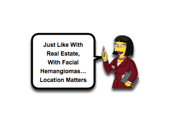
- Location matters! Facial hemangiomas are more likely to be associated with complications.
- Periorbital Hemangiomas and Bear Distribution Hemangiomas warrant a extra consideration!
- “Recurrent croup” may not be croup! Remain vigilant!
References
Darrow DH, Greene AK, Mancini AJ, Nopper AJ; SECTION ON DERMATOLOGY, SECTION ON OTOLARYNGOLOGY–HEAD AND NECK SURGERY, and SECTION ON PLASTIC SURGERY. Diagnosis and Management of Infantile Hemangioma. Pediatrics. 2015 Oct;136(4):e1060-104. PMID: 26416931. [PubMed] [Read by QxMD]
Satish V1, Bhat M2, Maganur PC3, Shah P4, Biradar V5. Capillary hemangioma in maxillary anterior region: a case report. Int J Clin Pediatr Dent. 2014 May;7(2):144-7. PMID: 25356016. [PubMed] [Read by QxMD]
O TM1, Scheuermann-Poley C, Tan M, Waner M. Distribution, clinical characteristics, and surgical treatment of lip infantile hemangiomas. JAMA Facial Plast Surg. 2013 Jul-Aug;15(4):292-304. PMID: 23752875. [PubMed] [Read by QxMD]
Sajan JA1, Tibesar R, Jabbour N, Lander T, Hilger P, Sidman J. Assessment of pulsed-dye laser therapy for pediatric cutaneous vascular anomalies. JAMA Facial Plast Surg. 2013 Nov-Dec;15(6):434-8. PMID: 24008312. [PubMed] [Read by QxMD]
Haggstrom AN1, Skillman S, Garzon MC, Drolet BA, Holland K, Matt B, McCuaig C, Metry DW, Morel K, Powell J, Frieden IJ. Clinical spectrum and risk of PHACE syndrome in cutaneous and airway hemangiomas. Arch Otolaryngol Head Neck Surg. 2011 Jul;137(7):680-7. PMID: 21768412. [PubMed] [Read by QxMD]
O-Lee TJ1, Messner A. Subglottic hemangioma. Otolaryngol Clin North Am. 2008 Oct;41(5):903-11, viii-ix. PMID: 18775341. [PubMed] [Read by QxMD]
Haggstrom AN1, Drolet BA, Baselga E, Chamlin SL, Garzon MC, Horii KA, Lucky AW, Mancini AJ, Metry DW, Newell B, Nopper AJ, Frieden IJ. Prospective study of infantile hemangiomas: clinical characteristics predicting complications and treatment. Pediatrics. 2006 Sep;118(3):882-7. PMID: 16950977. [PubMed] [Read by QxMD]
Kronenberg A1, Blei F, Ceisler E, Steele M, Furlan L, Kodsi S. Ocular and systemic manifestations of PHACES (Posterior fossa malformations, Hemangiomas, Arterial anomalies, Cardiac defects and coarctation of the Aorta, Eye abnormalities, and Sternal abnormalities or ventral developmental defects) syndrome. J AAPOS. 2005 Apr;9(2):169-73. PMID: 15838446. [PubMed] [Read by QxMD]
Williams EF 3rd1, Stanislaw P, Dupree M, Mourtzikos K, Mihm M, Shannon L. Hemangiomas in infants and children. An algorithm for intervention. Arch Facial Plast Surg. 2000 Apr-Jun;2(2):103-11. PMID: 10925435. [PubMed] [Read by QxMD]
Tanner JL1, Dechert MP, Frieden IJ. Growing up with a facial hemangioma: parent and child coping and adaptation. Pediatrics. 1998 Mar;101(3 Pt 1):446-52. PMID: 9481012. [PubMed] [Read by QxMD]
Orlow SJ1, Isakoff MS, Blei F. Increased risk of symptomatic hemangiomas of the airway in association with cutaneous hemangiomas in a “beard” distribution. J Pediatr. 1997 Oct;131(4):643-6. PMID: 9386676. [PubMed] [Read by QxMD]
Author

Thank you for pointing out that there are options for treatment when it comes to hemangioma. My sister is suffering from this and we are wanting to get it treated. I’ll have to look into finding the best treatment for her.
How effective is laser therapy in treatment of Facial Hemangioma.